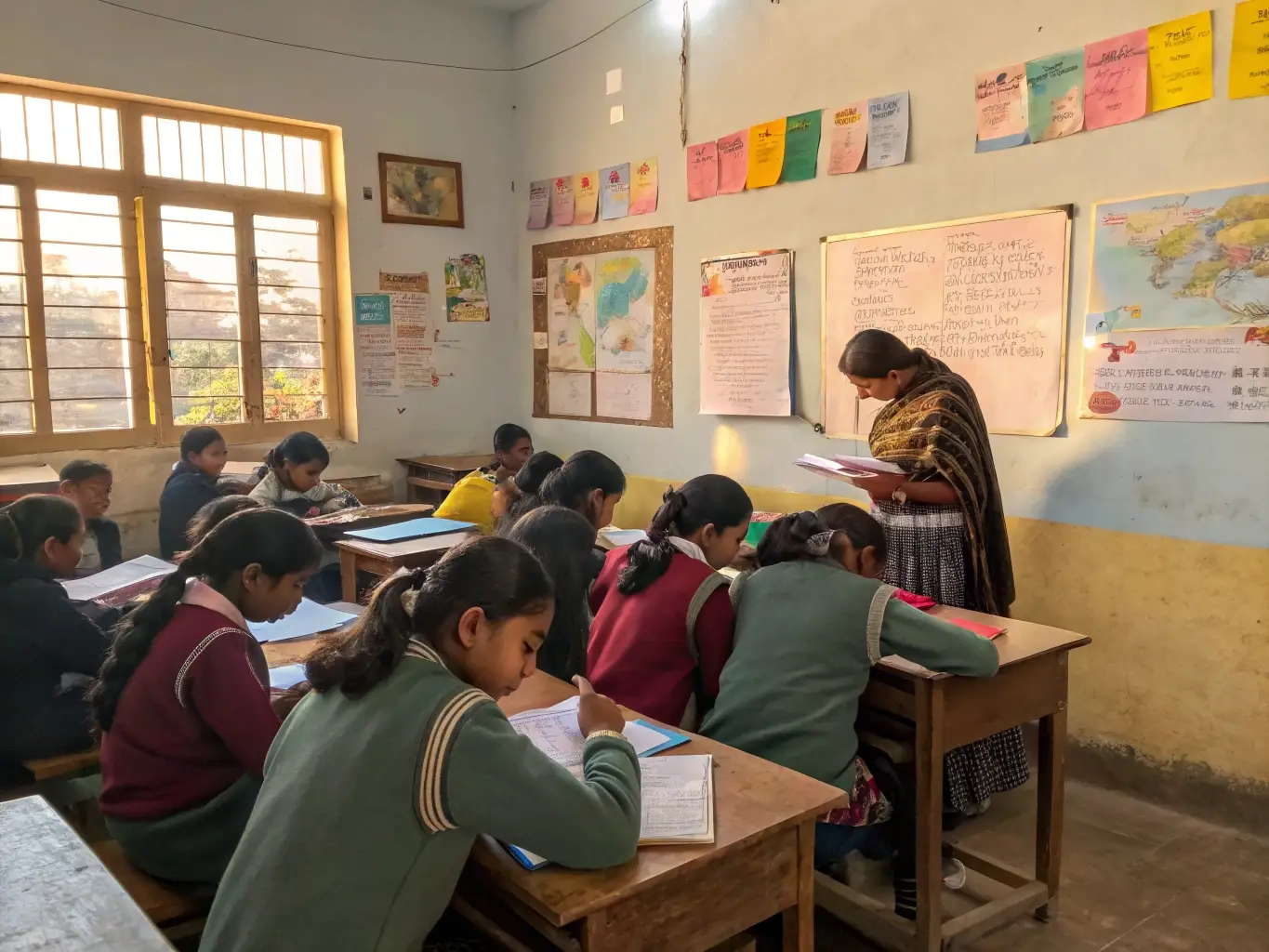
Chapter 6, Anatomy of Flowering Plants, takes students inside the plant body to study its tissues and tissue systems in detail. It builds on the tissue basics from earlier classes and explains meristematic and permanent tissues, the epidermal, ground and vascular tissue systems, and how these are organised differently in dicotyledonous and monocotyledonous roots, stems and leaves, along with an introduction to secondary growth.
Last Updated: September 23, 2026
Exercises
Q1. Draw illustrations to bring out the anatomical difference between (a) monocot root and dicot root (b) monocot stem and dicot stem — Since a hand-drawn diagram cannot be reproduced here, the anatomical differences that…
Since a hand-drawn diagram cannot be reproduced here, the anatomical differences that such a labelled diagram should bring out are summarised below.
(a) Monocot root vs Dicot root
| Feature | Dicot Root | Monocot Root |
|---|---|---|
| Cortex | Comparatively narrow, made up of parenchymatous cells with a distinct, single-layered endodermis showing casparian strips | Wide cortex, also parenchymatous, with a prominent endodermis |
| Xylem bundles (protoxylem groups) | Few, usually 2 to 6 (diarch to hexarch) | Many, usually more than six (polyarch) |
| Pith | Small or absent | Large and well developed, occupying the centre |
| Conjunctive tissue (between xylem and phloem) | Parenchymatous initially, but some cells become meristematic later to form vascular cambium | Remains permanently parenchymatous; never forms cambium |
| Secondary growth | Present, due to formation of vascular cambium and cork cambium | Absent |
| Root system | Tap root system | Fibrous/adventitious root system |
(b) Monocot stem vs Dicot stem
| Feature | Dicot Stem | Monocot Stem |
|---|---|---|
| Hypodermis | Collenchymatous (living, provides mechanical flexibility) | Sclerenchymatous (dead, lignified, provides rigidity) |
| Ground tissue | Differentiated into cortex, endodermis, pericycle, medullary rays and pith | Not differentiated into cortex and pith; a single continuous mass of parenchyma called ground tissue |
| Vascular bundles | Limited in number, arranged in a single ring; conjoint, collateral and open (cambium present between xylem and phloem) | Numerous, scattered throughout the ground tissue; conjoint, collateral and closed (no cambium) |
| Bundle sheath | Absent | Each vascular bundle is surrounded by a sclerenchymatous bundle sheath |
| Phloem parenchyma | Present | Absent |
| Xylem vessels | Usually arranged in a “Y” shape with distinct protoxylem and metaxylem | Arranged with a large, water-storing cavity near the protoxylem end |
| Secondary growth | Present (vascular cambium and cork cambium become active) | Absent |
Q2. Cut a transverse section of the young stem of a plant from your school garden and observe it under the microscope. How would you ascertain whether it is a monocot stem or a dicot stem? Give reasons — The identity of the stem can be established from the arrangement and structure of its…
The identity of the stem can be established from the arrangement and structure of its vascular bundles and ground tissue. If the section shows vascular bundles arranged in a single ring, each being conjoint, collateral and open (with a strip of cambium between xylem and phloem), and the ground tissue clearly differentiated into hypodermis (collenchyma), cortex, endodermis, pericycle and a central pith, the stem is a dicot stem. If instead the vascular bundles are numerous and scattered throughout an undifferentiated ground tissue, each bundle is conjoint, collateral and closed, surrounded by a sclerenchymatous bundle sheath, the hypodermis is sclerenchymatous, and the xylem vessels are arranged in a Y-shaped pattern with a large water cavity, the stem is a monocot stem. The single most reliable feature to check first is whether the vascular bundles are ringed (dicot) or scattered (monocot).
Q3. The transverse section of a plant material shows the following anatomical features – (a) the vascular bundles are conjoint, scattered and surrounded by a sclerenchymatous bundle sheath, (b) phloem parenchyma is absent. What will you identify it as? — This section should be identified as a monocot stem. Scattered vascular bundles enclosed…
This section should be identified as a monocot stem. Scattered vascular bundles enclosed by a sclerenchymatous bundle sheath and the absence of phloem parenchyma are diagnostic features found only in the stems of monocotyledonous plants; dicot stems have ringed, open vascular bundles without a sclerenchymatous bundle sheath, and phloem parenchyma is present in them.
Q4. What is stomatal apparatus? Explain the structure of stomata with a labelled diagram — The stomatal apparatus refers to the stomatal pore together with the two surrounding…
The stomatal apparatus refers to the stomatal pore together with the two surrounding guard cells and the adjacent epidermal cells, called subsidiary cells, which are often structurally modified. Stomata occur mainly in the epidermis of leaves and regulate transpiration and gaseous exchange.
Each stoma is bordered by two bean-shaped (kidney-shaped in dicots; dumb-bell shaped in many grasses/monocots) guard cells. The wall of the guard cell facing the pore (inner wall) is thick and rigid, while the wall away from the pore (outer wall) is thin and elastic. Guard cells are the only epidermal cells that contain chloroplasts. Changes in the turgor pressure of guard cells, caused by the inflow or outflow of water, alter their shape and thereby open or close the stomatal pore. When guard cells are turgid, the thin outer walls bulge out, forcing the thick inner walls apart and opening the pore; when they lose turgor, the pore closes. Subsidiary cells support the guard cells mechanically and help control this movement.
Q5. Name the three basic tissue systems in flowering plants. Give the tissue names under each system — The three basic tissue systems in flowering plants, first described by Sachs, are:
The three basic tissue systems in flowering plants, first described by Sachs, are:
1. Epidermal tissue system – comprises the epidermis (a single continuous outer layer, usually covered by a cuticle), stomata, and epidermal appendages such as trichomes (on the stem) and root hairs.
2. Ground tissue system – includes all tissue except the epidermis and vascular bundles: the simple tissues parenchyma, collenchyma and sclerenchyma. In roots and stems this forms the cortex, endodermis, pericycle, pith and medullary rays; in leaves it forms the mesophyll.
3. Vascular tissue system – consists of the complex tissues xylem (tracheids, vessels, xylem fibres, xylem parenchyma) and phloem (sieve tube elements, companion cells, phloem fibres, phloem parenchyma), organised into vascular bundles.
Q6. How is the study of plant anatomy useful to us? — Plant anatomy, the study of the internal structure of plants, has several practical and…
Plant anatomy, the study of the internal structure of plants, has several practical and academic applications:
It helps in understanding the structural adaptations plants develop in response to different climatic and habitat conditions (for example, xerophytic or hydrophytic modifications). It is used to distinguish monocots, dicots and gymnosperms on the basis of internal structure, which is valuable in plant classification and solving taxonomic problems. It assists in judging the quality and strength of wood and commercial fibres (such as jute and flax), helping identify superior versus substandard timber for industrial use. It also supports pharmacognosy — establishing the correct identity and purity of plant-derived drug material — and helps trace evolutionary relationships (homology and analogy) among plant groups.
Q7. Describe the internal structure of a dorsiventral leaf with the help of a labelled diagram — A dorsiventral (bifacial) leaf, typical of dicotyledons, is oriented horizontally and…
A dorsiventral (bifacial) leaf, typical of dicotyledons, is oriented horizontally and shows structural differences between its upper and lower surfaces. In transverse section it shows the following layers:
Upper (adaxial) epidermis – a single outer layer of parenchymatous cells covered by a cuticle that reduces water loss; it is generally without chloroplasts and has few or no stomata.
Lower (abaxial) epidermis – similar in structure to the upper epidermis, but it bears a greater number of stomata, through which most gaseous exchange and transpiration occur.
Mesophyll – the ground tissue between the two epidermal layers, made of thin-walled, chloroplast-containing parenchyma, differentiated into two zones: (i) palisade parenchyma — tall, columnar cells arranged compactly (usually in one to three rows) just beneath the upper epidermis, containing most of the chloroplasts and carrying out the bulk of photosynthesis; and (ii) spongy parenchyma — rounded or oval, loosely arranged cells with large intercellular spaces, located towards the lower epidermis, which facilitate gaseous exchange.
Vascular bundles – found within the mesophyll, running through the veins; each is conjoint, collateral and closed, with xylem oriented towards the upper epidermis and phloem towards the lower epidermis, and is surrounded by a parenchymatous bundle sheath. The vascular bundle in the midrib region is the largest.
Class 11 Biology Chapter 6 – Notes and Extra Questions
The current (2023 rationalised) NCERT exercise for this chapter has been trimmed down to just 7 questions, focused mainly on the anatomical comparison of monocot and dicot organs, stomatal structure, the three tissue systems, and dorsiventral leaf anatomy. Five questions that appeared in the older, pre-2023 edition — on the location/function of meristems, the statement that cork cambium forms the cork, a full schematic explanation of secondary growth in woody stems, why xylem and phloem are called complex tissues, and periderm formation — have been officially dropped along with large parts of the “Meristematic Tissues” and “Secondary Growth” sections of the chapter text (including vascular cambium activity, spring/autumn wood, heartwood/sapwood, cork cambium and secondary growth in roots). While these dropped topics are no longer examined directly through the textbook exercise, students should still know the terms parenchyma, collenchyma, sclerenchyma, casparian strips, bulliform cells and the basic idea of secondary growth, since they are frequently tested in school unit tests, board pre-boards and competitive exams such as NEET even when not part of the current CBSE textbook exercise.
- Chapter 1: The Living World – Free PDF Download
- Chapter 2: Biological Classification – Free PDF Download
- Chapter 3: Plant Kingdom – Free PDF Download
- Chapter 4: Animal Kingdom – Free PDF Download
- Chapter 5: Morphology of Flowering Plants – Free PDF Download
- Chapter 7: Structural Organisation in Animals – Free PDF Download
- Chapter 8: Cell - The Unit of Life – Free PDF Download
- Chapter 9: Biomolecules – Free PDF Download
- Chapter 10: Cell Cycle and Cell Division – Free PDF Download
- Chapter 11: Photosynthesis in Higher Plants – Free PDF Download
- Chapter 12: Respiration in Plants – Free PDF Download
- Chapter 13: Plant Growth and Development – Free PDF Download
- Chapter 14: Breathing and Exchange of Gases – Free PDF Download
- Chapter 15: Body Fluids and Circulation – Free PDF Download
- Chapter 16: Excretory Products and their Elimination – Free PDF Download
- Chapter 17: Locomotion and Movement – Free PDF Download
- Chapter 18: Neural Control and Coordination – Free PDF Download
- Chapter 19: Chemical Coordination and Integration – Free PDF Download
Frequently Asked Questions
What is the difference between simple and complex tissues in plants?
Simple tissues (parenchyma, collenchyma, sclerenchyma) are made up of only one type of cell that is structurally and functionally similar throughout. Complex tissues (xylem and phloem) are made up of more than one type of cell that work together as a single functional unit — for example, xylem includes tracheids, vessels, xylem fibres and xylem parenchyma, all cooperating in water conduction, while phloem includes sieve tube elements, companion cells, phloem fibres and phloem parenchyma, cooperating in food transport.
Why does a dicot stem show secondary growth while a monocot stem generally does not?
Secondary growth occurs because of lateral meristems — the vascular cambium and cork cambium. Dicot stems have open vascular bundles, meaning a strip of cambium is already present between the xylem and phloem; this fascicular cambium connects with interfascicular cambium (formed from medullary ray cells) to form a complete ring capable of adding secondary xylem and phloem. Monocot stems have closed vascular bundles with no cambium between the xylem and phloem, so they lack the meristematic tissue needed to increase in girth, and therefore almost never show secondary growth.
What are casparian strips and where are they found?
Casparian strips are band-like thickenings of suberin (a waxy, waterproofing substance) deposited on the radial and transverse walls of the endodermal cells that surround the vascular cylinder of a root. They force water and dissolved minerals travelling through the cell walls (the apoplast pathway) to enter the endodermal cell membrane and cytoplasm (the symplast pathway) before reaching the xylem, giving the plant control over which substances are absorbed into the vascular tissue.
What are bulliform cells and what is their function in monocot leaves?
Bulliform cells are large, empty, thin-walled, colourless cells found on the upper (adaxial) epidermis of many monocot leaves, especially grasses, usually occurring in groups along the veins. When water is plentiful, they remain turgid and the leaf stays flat and expanded. During water stress, they lose turgor and shrink, causing the leaf to roll or fold inward, which reduces the exposed surface area and minimises water loss through transpiration.
Recommended: Buy the Printed NCERT Class 11 Biology Book
Contains Amazon affiliate links.
If you’d like a printed copy alongside the PDF, here’s a verified option:
4.1 out of 5 stars (973 ratings) · Rs. 183
Price and availability may change on Amazon. As an Amazon Associate, ncertbooks.org earns from qualifying purchases.